The James L. Voss Veterinary Teaching Hospital celebrated the opening of the Lucy & Family PET/CT Suite last week. This suite houses a state-of-the-art Siemens PET/CT scanner that adds to the hospital’s advanced diagnostic imaging tools and brings Colorado State University one step closer to being a full Siemens campus, the industry leader for advanced imaging.
The Lucy & Family PET/CT Suite is 1375 square feet, complete with the machine, a holding area, anesthesia and recovery space, as well as a control room. It was designed with patients in mind – easing the stress of the procedure and improving workflows to streamline the process.
This space previously held the prior PET/CT machine; it was the first dedicated veterinary PET/CT scanner installed anywhere in the world. The previous machine was used for many years to diagnose both large and small animals. With the retirement of the previous machine, the space has been completely renovated and upgraded with the latest technology.
A PET/CT is a diagnostic imaging tool that allows clinicians to evaluate how their patients’ organs are functioning through the use of short-acting radioactive compounds, called radiotracers. These radiotracers accumulate in regions of the body with atypical metabolic function or cellular changes, such as diseased or cancerous tissue. These regions are then highlighted during the scan to show which regions of the body are functioning abnormally and may be affected by cancer or other disease states.
When a PET and CT scan are combined, clinicians can get detailed information about both their patients’ anatomy and physiology, or organ function, to get a full 3D picture of the patient’s internal health. By combining the information, they can detect regions of disease earlier and more accurately. These scans allow us to plan highly specific and targeted treatment options for patients.
The Imaging Core at CSU
The PET/CT machine is just the latest addition to the hospital’s deep diagnostic capabilities. The Lucy & Friends CT Suite was opened in 2020 and boasts an advanced CT machine.
A CT scan is an important imaging tool used for both diagnosing disease and treatment planning for pets and humans alike. CT scans are performed by rotating X-rays around a patient’s body and using computer software to create cross-sectional images of the body in slices. This provides a comprehensive 3D look inside a patient’s body by eliminating the superimposition of organs and tissues that occurs with regular X-rays. These high-quality images give us detailed information to help diagnose cancer, injury, heart disease, or other diseases and simultaneously help plan surgery, radiation, or other treatment options.
Since the Lucy & Friends Suite opened, the machine has been used to scan 4,022 patients. Of those scans, 2,388 had radiation planning scans. Radiation planning scans create special images that allow us to create highly targeted radiation treatment plans to eradicate cancerous growths.
The CT scanner has been used to obtain diagnostic imaging to diagnose a variety of issues in nearly every specialty at the VTH including cardiology, dentistry, exotics, neurology, oncology, orthopedic surgery, orthopedic medicine and mobility, urgent and critical care, and small animal medicine. Specific reasons to use this machine include planning a cardiac valve replacement, repair, or stent through an interventional approach, identifying a specific anatomic location for a tumor, and planning for radiation therapy. We also use these machines to help us understand which structures are involved in lameness.
For all cases, we use the scans to gather diagnostic information to determine the best course of treatment.
The Impact
These machines have an enormous impact on the hospital, and ultimately on patient care. The flexibility to run CT or PET/CT scans gives our faculty and students the option to plan diagnostic studies that meet their patient’s needs. The James L. Voss Veterinary Teaching Hospital was one of the first veterinary practices to offer patients PET scans.
Having two state-of-the-art machines doubles the hospital’s bandwidth and allows for more patients to receive advanced scans as part of their diagnostic work-up and treatment planning. In addition, the Lucy and Family suite includes a satellite anesthesia area adjacent to the advanced imaging suites.
Furthermore, both machines are fast CT scanners. Patients can be in and out of the machine with a full scan in less than two minutes. This means that animals will be under anesthesia for significantly less time and results can be processed much more quickly.
PET/CTs and CT scans are used by a variety of services across the hospital. Oncology uses the machine to scan for cancerous tumors and monitor growth and activity. Neurology uses the scanner to map brain activity and diagnose neurological disorders. The Mobility and PT departments rely on this imaging tool to assess joint, muscle, and tissue damage. Cardiology depends heavily on CT scanners to plan for surgeries and interventional approaches such as stent placement and valve replacements.
With the capabilities of both machines, we aim to increase CT scans by as much as 50%. This diagnostic modality is standard of care in human medicine and we aim to do the same in veterinary care.
“The original PET/CT put CSU at the forefront of cancer imaging in veterinary medicine. The two new machines, the dual-energy CT and the new PET/CT again put CSU at the leading edge of veterinary imaging. The type of images they provide and the speed at which the images are acquired help the entire hospital provide world-class care to our patients and their families,” said Dr. Elissa Randall, Professor of Veterinary Diagnostic Imaging at Colorado State University.
Lucy’s Legacy

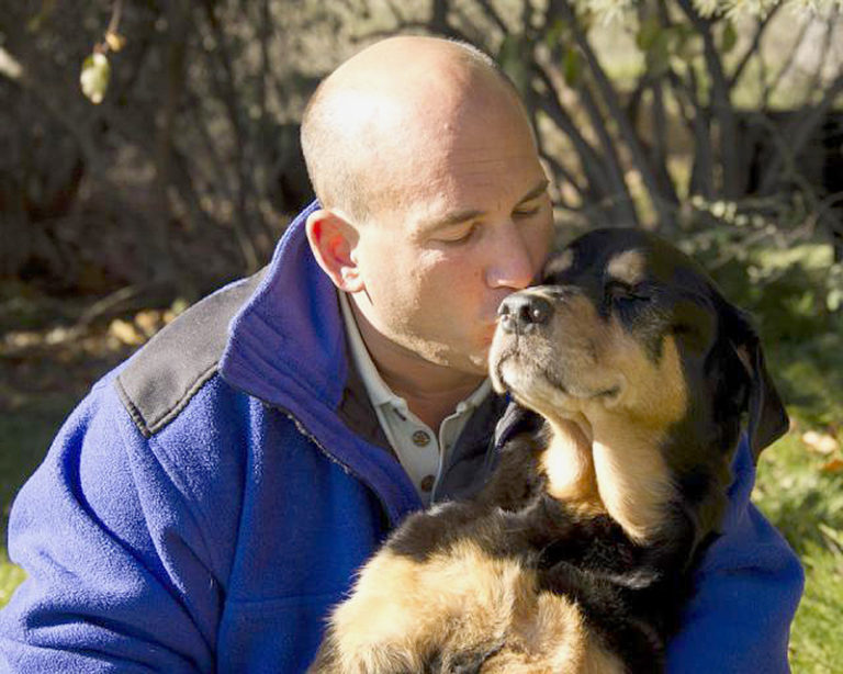
The imaging Suites are named after a special dog named Lucy and she’s created quite a legacy here at Colorado State University.
Jeffrey Neu brought his beloved dog, Lucy, to CSU in 2010. The hospital’s original dedicated PET/CT scanner, unusual in veterinary medicine, inspired Jeff to bring Lucy from California. She was the first patient to receive serial PET/CT scans to monitor her disease, the standard of care in human oncology.
Lucy had osteosarcoma; at CSU, she received a PET scan, which allowed early detection of metastasized bone cancer. The scans gave her clinicians the information necessary to make an informed treatment plan. She underwent chemotherapy and precisely targeted radiation; the treatments extended Lucy’s life and improved her quality of life until she succumbed to the disease.
Jeffrey and Tiffany Neu have made both the Lucy & Friends CT and the Lucy & Family PET/CT Suites possible with their generous donations. The Lucy & Friends room honors the working search and rescue dogs Jeff has worked with, while the Lucy & Family suite honors all of the Neu family pets.
“My dreams are finally coming true – all patients who visit Colorado State University will have access to the amazing diagnostic imaging that Lucy received during her first visit in 2010. With two fast scanners at CSU, every patient who needs a scan can get one,” said Jeffrey Neu. “This allows not only for better diagnostic information but also provides better care for pets. Furthermore, a second fast CT and a state-of-the-art PET/CT scanner in the hospital provides greater research opportunities. There is potential to study different isotopes and I’m eager to see what researchers will find. Ultimately these machines will help pave the way for better treatments for animals now and in the future.”
“We simply could not do what we do without the support of our friends,” said Dr. Susan Lana, Interim Director of the Flint Animal Cancer Center. “The Neus have continually made advancements in veterinary medicine possible, and for that we’re grateful.”
These machines allow us to provide the best possible care for our patients. They also give our students advanced training so that they can become the veterinarians of tomorrow. This is all possible thanks to a special dog named Lucy.